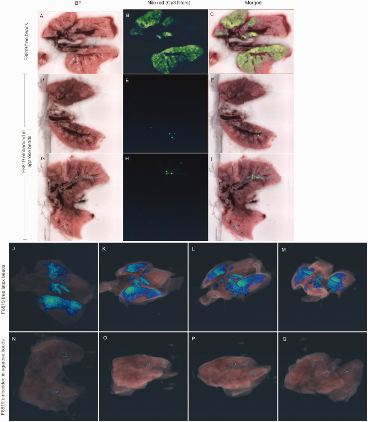
Figure 3.
Imaging whole lungs shows different distribution of fluorescent particles when embedded in agarose compared with free latex beads. Cross-section images from lungs of a mouse administered a suspension of fluorescent F8819 latex particles (a)–(c). Two different cross-sections from lungs of a mouse administered fluorescent F8819 latex particles embedded in agarose beads (d)–(i). (a), (d), and (g) are brightfield; (b), (e), and (h) show Nile red fluorescence, and (c), (f), and (i) are the merged images. Serial images, such as (f) and (i), are compiled to reconstruct a 3D view of the lungs. (j)–(m) show different rotational views of the lungs of a mouse given the F8819 particle suspension, while (n)–(q) show the distribution of F8819 particles embedded in agarose.

