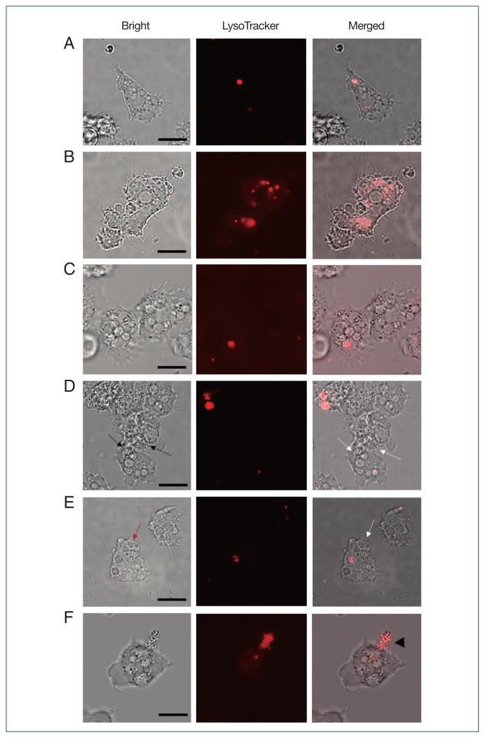
Fig. 4.
LysoTracker staining of siRNA-transfected Acanthamoeba containing E. coli. The Acanthamoeba that did and did not undergo siRNA transfection were allowed to ingest E. coli for 12 h, and those containing E. coli were stained with LysoTracker. (A) A. castellanii control, (B) A. castellanii containing E. coli, (C) siRNA-transfected A. castellanii control, (D) ACA1_077100 siRNA-transfected A. castellanii containing E. coli, (E) ACA1_175060 siRNA-transfected A. castellanii containing E. coli, and (F) AFD36229.1 siRNA-transfected A. castellanii containing E. coli. Black arrows, E. coli in the cytoplasm of Acanthamoeba. Red arrow, E. coli in the phagosomes of Acanthamoeba; White arrows, E. coli in Acanthamoeba cytoplasm or phagosomes without lysosomes. Black arrowhead, E. coli that burst out of Acanthamoeba. Bar=10 μm.

