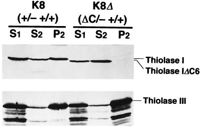
FIG. 7.
Subcellular distribution of the wild-type Thiolase I [K8 (+/− +/+)] and C-terminus-deleted Thiolase I [K8Δ (ΔC/− +/+)]. Cells grown on n-alkanes were harvested at mid-logarithmic phase, lysed to protoplast, homogenized, and separated to nuclear and postnuclear fractions (51). S1, S2, and P2 represent postnuclear supernatant, cytoplasm/microsome, and organelle fractions, respectively. Protein (20 μg) from each fraction was run on gels. Thiolases were detected with anti-Ps-Thiolase I (upper panel) and anti-Thiolase III (lower panel) antisera. Thiolase IΔC6, C-terminus-truncated Thiolase I.

