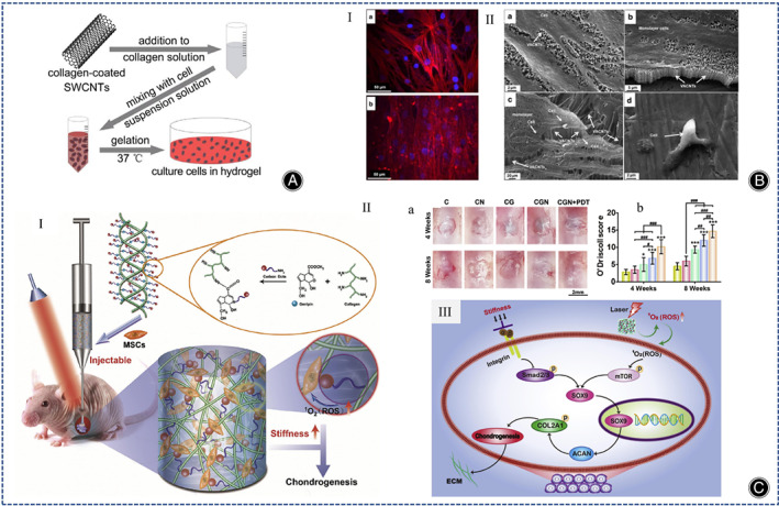
Fig. 6.

(A) Illustration of the synthesis of SWCNT/collagen composite hydrogels. (Figures are adapted with permission from Mao et al. 72 ) (B) (I) Fluorescence microscopy images of Actin (red) and DAPI (blue) of human chondrocytes (scale bar is 50μm). (a) Chondrocytes in superhydrophilic VACNT; (b) chondrocytes on the membrane. (II) SEM images of chondrocytes after 5 days of culture. (a, b) Characterization of human chondrocytes in superhydrophilic VACNT membranes and (c) VACNT/PMMA membranes; (d) chondrocytes on membranes with arrows pointing to chondrocytes and VACNTs. (Figures are adapted with permission from Antonioli et al. 75 ) (C) (I) Schematic diagram of the preparation process of collagen‐genipin‐carbon dot (CGN) nanocomposite hydrogel. (II) PDT promotes cartilage regeneration after CGN implantation. (a, b) Macroscopic images of cartilage tissue engineering (a) and O'Driscoll score (b). (Scale bar is 2 mm) (C represents collagen, CN represents collagen mixed with CD NPs, CG represents collagen cross‐linked with genipin, CGN represents collagen cross‐linked with genipin and CD NPs, and CGN + PDT represents CGN after 3 min of 808 nm laser irradiation at a power density of 5.6 mW/cm2). (III) Schematic diagram of the signaling pathways of mechanotransduction and laser irradiation. (Figures are adapted with permission from Lu et al. 80 ).
