Fig. 6.
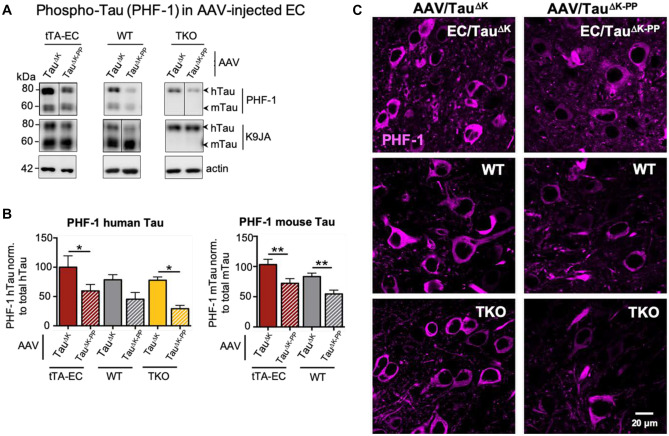
Increased Tau phosphorylation (pS396/pS404) in the EC of AAV/TauK injected mice. A Representative western blot analysis of Tau phosphoprylated at residues Ser396/Ser404 (PHF-1) and of total Tau (K9JA) in EC lysates from tTA-EC mice (EC/TauΔK or EC/TauΔK−PP, lanes 1, 2), WT (lanes 3, 4), and TKO mice (lanes 5, 6), each injected with either AAV/TauΔK (lanes 1, 3, 5) or AAV/TauΔK−PP (lanes 2, 4, 6) at 3 months p.i. Lanes 1, 2: [tTA-EC + AAV]-induced expression of pro- and anti-aggregant human Tau yields pronounced bands of mTau and hTau, both phosphorylated at the PHF-1 epitope. Lanes 3, 4: [WT + AAV] mice showed pronounced mTau, lesser hTau, but pronounced PHF-1 reactivity only in pro-aggregant hTau. Lanes 5, 6: [TKO + AAV] mice showed only hTau and pronounced PHF-1 reactivity only in the pro-aggregant mice (lane 5, top). The data illustrate that pro-aggregant hTau is more prone to phosphorylation by PHF-1. B Differential quantification of phosphorylated human and mouse Tau shows that AAV/TauΔK injected mice have generally higher (~30–40%) PHF-1 Tau levels (bars 1, 3, 5) than AAV/TauΔK−PP injected animals (bars 2, 4, 6). One-way ANOVA with post-hoc uncorrected Fisher’s LSD test for multiple comparisons. *p < 0.05; **p < 0.01. Data shown as mean ± SEM. C Immunolabeling with PHF-1 antibody detects higher amounts of phospho-Tau (pS396/pS404) in brain sections from tTA-EC, WT, and TKO mice injected with AAV/TauΔK compared to AAV/TauΔK−PP injected animals. Scale bar: 20 µm

