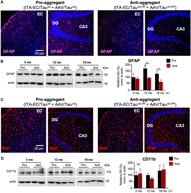
Fig. 8.
Density of astrocytes and microglia in pro-aggregant and anti-aggregant neuropsin mice. A Immunolabeling of astrocytes (GFAP) in brain sections suggests an increased number and fluorescent intensity of astrocytes, reminiscent of a mild astrogliosis, in the hippocampal formation (CA3 and around the DG granule cell layer) of pro-aggregant AAV/TauΔK-injected tTA-EC/TauΔK mice compared to anti-aggregant AAV/TauΔK−PP-injected tTA-EC/TauΔK−PP mice. Scale bar: 50 μm. B Western blot analysis of EC lysates shows that GFAP increased in the pro-aggregant mice from 3 to 12 months p.i. (p = 0.0227) and was significantly higher (~40%) in AAV/TauΔK-injected tTA-EC/TauΔK mice compared to AAV/TauΔK−PP-injected tTA-EC/TauΔK−PP mice at longer post-injection times of 12 (p = 0.0157) and 18 (p = 0.0373) months. Two-way ANOVA; with post-hoc uncorrected Fisher’s LSD test. Data shown as mean ± SEM. C No differences were observed in the expression of Iba1 between the EC and hippocampal region in the pro-aggregant mice. Regarding Iba1 expression, no differences were observed between pro- and anti-aggregant mice, as well as between the EC and hippocampal region. D WB quantification of the levels of the microglial marker CD11b confirmed the absence of differences between pro- and anti-aggregant mice in terms of microglia at the time points analyzed (3, 12 and 18 months p.i.). Data shown as mean ± SEM

