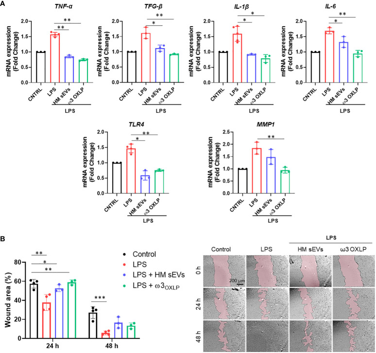
Figure 4.
HM sEVs and ω3 OXLP prevent LPS-induced fibrosis. (A) Expression levels of TNF-α, TGF-β, IL-1β, IL-6, TLR4 and MMP1 quantified by RT-qPCR in fibroblasts stimulated with lipopolysaccharides (LPS) and/or treated with 7.5 µg/mL sEVs or 0.5 nM of each of the three ω3 OXLP. Unstimulated fibroblasts were used as controls. The expression level of the target gene in each sample was normalized to GAPDH expression. represented as mean percentage ± SD. (B) Quantification of fibroblast wound closure at 24 and 48 h. Data were normalized to initial wound area and are represented as mean percentage ± SD. Representative brightfield images of wound healing assay at different times (0, 24 and 48 h) after wound generation on a monolayer fibroblast culture stimulated with LPS alone or treated with 7.5 µg/mL sEVs or 0.5 nM of each of the three ω3 OXLP. Images were taken at 10× magnification. Scale bar: 200 µm. Experiments were performed in triplicate. One-way ANOVA was used for statistical analysis. *p< 0.05, **p< 0.01, ***p< 0.001.

