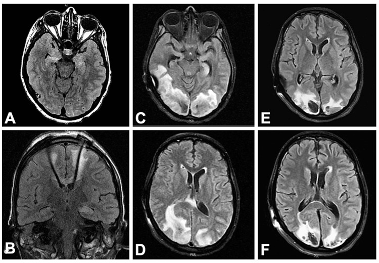
Figure 2. Evolution of neuroimaging.
A: T2 hyperintensity of bilateral mesial temporal lobes on admission; B: Placement of deep brain stimulator with electrode tips terminating in anterior thalami nuclei; C,D: Confluent posterior predominant, asymmetric subcortical and deep white matter hyperintensity and edema consistent with PRES; E,F: Repeat imaging two weeks later showing resolving prior T2 lesions and development of intraparenchymal hematomas

