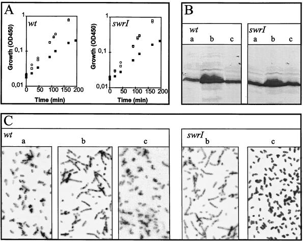
FIG. 2.
Analysis of S. liquefaciens strains harboring plasmid pMG600. (A) Growth; (B) flagellar content; (C) cell shape. Cells were grown in liquid cultures with no addition (□ and a), 0.5 mM IPTG (▪ and b), and 20 μM BHL (○ and c). Cells were harvested from the growing cultures at an optical density at 450 nm (OD450) of 0.25 for analysis in panels B and C. (B) Aliquots (50 μl) were heat denatured in sodium dodecyl sulfate-containing sample buffer, and the proteins were then separated by means of a standard sodium dodecyl sulfate-PAGE procedure, transferred to an Immobilon-P membrane (Millipore), and subjected to Western blotting analysis with rabbit antibodies directed against S. liquefaciens flagellar protein. Following binding of secondary alkaline phosphatase-labeled anti-rabbit immunoglobulin G, detection was performed with p-nitroblue tetrazolium chloride and 5-bromo-4-chloro-3-indolylphosphate. (C) Microscopic inspection of cell shape with a 50× long-working-distance objective.

