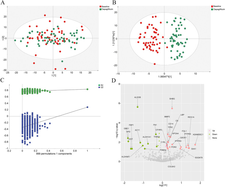
Fig. 1.
The differential protein expression analysis in the T2D patients at the baseline and after dapagliflozin treatment. A PCA score plot of proteomic data in the T2D patients at the baseline (red dots) and after dapagliflozin treatment (green dots). The cumulative fitness (R2 value) of the PCA model was 0.558. The t [1] and t [2] values in the figures represent the scores of each sample in principal components 1 and 2, respectively. Each dot on the plot represents a sample in the corresponding group. B OPLS-DA score plot of proteomic data in the T2D patients at the baseline (red dots) and after dapagliflozin treatment (green dots) (R2Y = 0.841, Q2 = 0.285). Each dot on the plot represents a sample in the corresponding group. C Permutation test of OPLS-DA (R2 intercept = 0.773, Q2 intercept = − 0.356). D Volcano plots of the differentially abundant proteins in the T2D patients after dapagliflozin treatment. The horizontal axis reflects fold change of the proteins after Log2 logarithmic conversion, and the vertical axis reflects FDR corrected p-value (q-value) after − Log10 logarithmic conversion. In the figure, the pink points indicate significantly differentially increased proteins, and green points indicate significantly differentially decreased proteins

