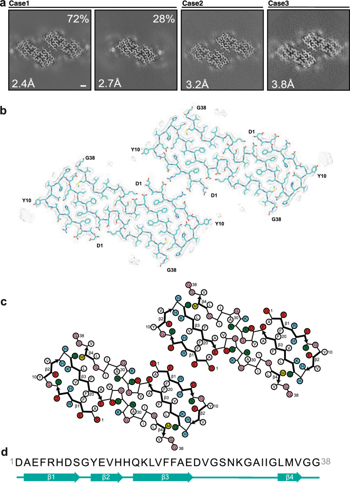
Fig. 2.
Structures of Aβ40 filaments from leptomeninges extracted using sarkosyl. a Cross-sections of Aβ40 filaments from cases 1–3 perpendicular to the helical axis, with a projected thickness of approximately one rung. Percentages of filaments are shown on the top right. The resolutions of the cryo-EM maps are indicated on the bottom left. Filaments were made of one (type 1) or two (type 2) pairs of protofilaments. Scale bar, 1 nm. b Cryo-EM density maps (transparent grey) and atomic model (cyan) of type 2 Aβ40 filaments with two pairs of protofilaments. c Schematic of the structure shown in panel b. Negatively charged residues are shown in red, positively charged residues in blue, polar residues in green, non-polar residues in white, sulphur-containing residues in yellow and glycines in pink. Thick connecting lines with arrowheads indicate β-strands. d Amino acid sequence of the core of Aβ40 protofilaments, which extends from D1 to G38. Beta-strands (β1–β4) are indicated as thick arrows

