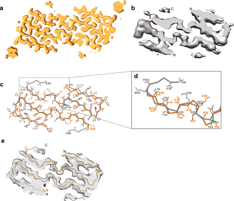
Fig. 5.
Comparison of the maps and models of Aβ40 filaments described here with those of Kollmer et al. [10]. a Map of Aβ40 filaments from leptomeninges with two protofilaments extracted using sarkosyl (present work; in orange). N- and C-termini are indicated. b Map of Aβ40 filaments from leptomeninges with two protofilaments extracted using a water-based method (previous work; in grey). N- and C-termini are indicated. c Atomic models of Aβ40 filaments for the maps shown in panels a and b (present work in orange; previous work in grey). The blue arrow points to V24, from which the two models differ in the register of their main chains. d Zoomed-in view of the structures in panel c. e Atomic model from present work (orange) and from previous work (grey) fitted into the 4.4 Å map from the previous work

