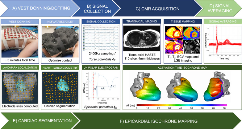
Fig. 3.
Step by step CMR-ECGI workflow. a The participant lies in the supine position for a 5-min recording at rest and an inflatable gilet is worn over the electrode vest to ensure good skin–electrode contact. b 256 ECG signals (torso potentials ϕT) are collected from the dry textile electrodes and amplified using the g.HIamp at a sampling frequency of 2400 Hz and processed using g.Recorder. c CMR scanning is performed at 3T to include T1 mapping, T2 mapping, LGE and ECV and transaxial HASTE used for cardiac segmentation and landmark localisation. d ECG signals are post processed using in-house Matlab software. e Heart-torso geometry (A) is generated from the HASTE via the reconstruction of individual cardiac meshes and imputation of the electrode markers using Amira software. f Heart-torso geometry (A) is combined with the torso potentials (ϕT) to solve the ‘inverse solution’ of ECGI (ϕT = AϕE) to reconstruct epicardial potentials (ϕE) at 1000 individual cardiac sites which are used to compute AT, RT and ARI per site and generate isochrone maps of cardiac excitation. In this example of a 75-year-old participant the earliest site of activation is in the posterolateral lateral wall of the LV. Ao aorta, ARI activation recovery interval, ECV extracellular volume, F frequency, LA left atria, LGE late gadolinium enhancement, LV left ventricle, mV millivolts, ms milliseconds, PA pulmonary artery, RA right ventricle, RV right ventricle, UEG unipolar electrogram. Other abbreviations as in Fig. 1

