Fig. 4.
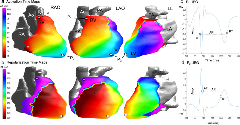
Epicardial isochrone maps. a AT maps in a healthy 75 year old male participant from the MyoFit46 cohort oriented here to mimic the 3 classic angiographic views in normal sinus rhythm. The earliest epicardial breakthrough (*) occurs in the basal RV in this participant. b RT maps in the same participant showing the wave of epicardial repolarization travelling from apex to base. c Reconstructed epicardial UEG for P1 which represents one node out of 1000 generated for individual cardiac sites—it highlights a negative QRS and T wave signal at the cardiac base. d Reconstructed epicardial UEG for P2 highlighting a positive QRS and T-wave signal at the cardiac apex. Each UEG is used to compute individual AT, RT, ARI and amplitudes per node which can then be averaged across all cardiac sites for the purposes of analysis. Amp amplitude, LAO left anterior oblique, LL left lateral, P1 point 1, P2 point 2, RAO right anterior oblique. Other abbreviations as in Fig. 1

