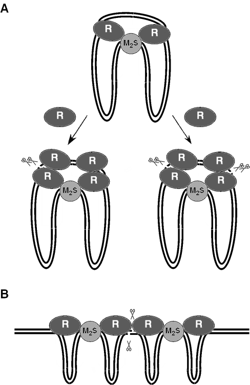
Figure 5.
Proposed models for DNA duplex breakage by Type I restriction enzymes on circular (A) and linear (B) DNA substrates. For more explanation see Discussion. DNA is represented as two black lines. The methylase (M2S1) and HsdR (R) subunits are represented as light gray and dark gray ovals. The pairs of scissors indicate the position of DNA double-strand break.

