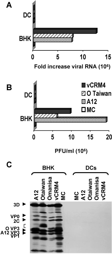
FIG. 1.
Analysis of FMDV replication in DC. DC were infected with FMDV at an MOI of 1 and cultured in complete medium for 0, 3, or 20 h. Samples of RNA were collected 0 and 3 h after virus adsorption. Culture supernatants were collected 0 and 24 h after adsorption for FMDV plaque titration. The permissive BHK cell line was infected under the same conditions as the positive control for virus replication. (A) Viral RNA levels measured by real-time PCR, with 18S rRNA as an internal control. The data are expressed as fold increases in the levels of FMDV RNA over the levels obtained 0 h after virus adsorption. (B) Standard virus plaque titrations performed with BHK cell monolayers. The data are expressed in PFU per milliliter of culture supernatant. The data shown here are averages of two different experiments. (C) Immunoprecipitation analysis of FMDV proteins in BHK cells and DC.

