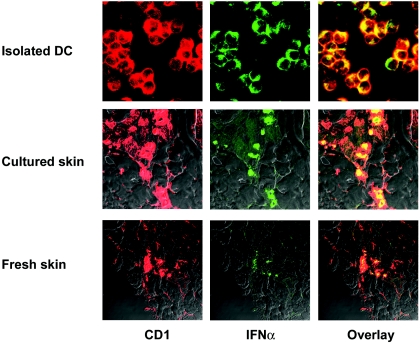
FIG. 7.
Confocal microscopy analysis of IFN-α protein expression by DC. Staining was performed to detect the expression of CD1 (red) and IFN-α (green) by isolated DC or in situ DC in skin sections. Skin layers were collected from healthy pigs. Frozen sections were prepared from samples collected immediately after the euthanasia of pigs (fresh skin) or after overnight culture (cultured skin) for DC isolation and were fixed in acetone. The data shown are representative of six different experiments.

