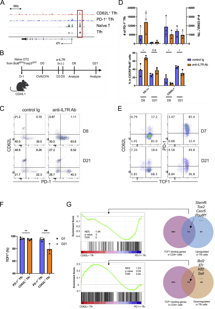
Figure S5.
IL7R signaling regulates CD62L+ Tfh cell development. (A) Mean ATAC-seq coverage at the Il7r gene locus in CD62L+ Tfh cell, PD1+ Tfh, naïve T cell, and in vitro induced Tfh-like cells. (B) Schematic diagram of IL-7R blocking antibody treatment in mice adoptively transferred with naïve OT-II+ T cells. (C) Representative flow cytometry plots of CD62L and PD-1 expression in the transferred CD45.2+ OT-II+ CD4+CXCR5+Bcl6+ Tfh cells from control and IL-7R blocking antibody-treated recipient mice. (D) Statistical analysis of PD-1+ and CD62L+ cell numbers (upper) and percentages (bottom) in Tfh cells of control and IL-7R blocking antibody-treated recipient mice at indicated time points. (E and F) B6 mice were subcutaneously immunized with KLH and analyzed on day 8 and 21 after immunization. (E) Representative flow cytometry staining of TCF1 versus CD62L and PD-1 in CD4+CD44hiCXCR5+Bcl6+ cells. (F) TCF1+ percentages in PD-1+ and CD62L+ Tfh cells. (G) GSEA indicated the genes upregulated and downregulated by TCF7 in CD62L+ Tfh cells and PD1+ Tfh cells. Data of B–F represent two independent experiments. Data are shown as mean ± SD; two-tailed t test; *, P < 0.05; **, P < 0.01; ns, no significance.

