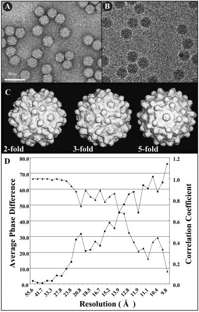
FIG. 1.
Cryo-EM reconstruction of wild-type AAV4. Negatively stained (A) and frozen (B) micrographs of wt AAV4 particles are shown. The majority of the particles were boxed from carbon film as shown in panel B. Bar = 50 nm. (C) Surface-shaded cryo-EM reconstructed AAV4 images (Purdue image format maps [pifmaps]) viewed down the icosahedral twofold (left), threefold (center), and fivefold (right) axes. The pifmaps were generated from 2D projections by use of the EM3DR subroutine in the Purdue suite of EM programs (6). (D) AAV4 map resolution (Å) determination. The resolution was determined to be where the average phase difference dropped below 50% or the correlation coefficient dropped below 0.5 (6).

