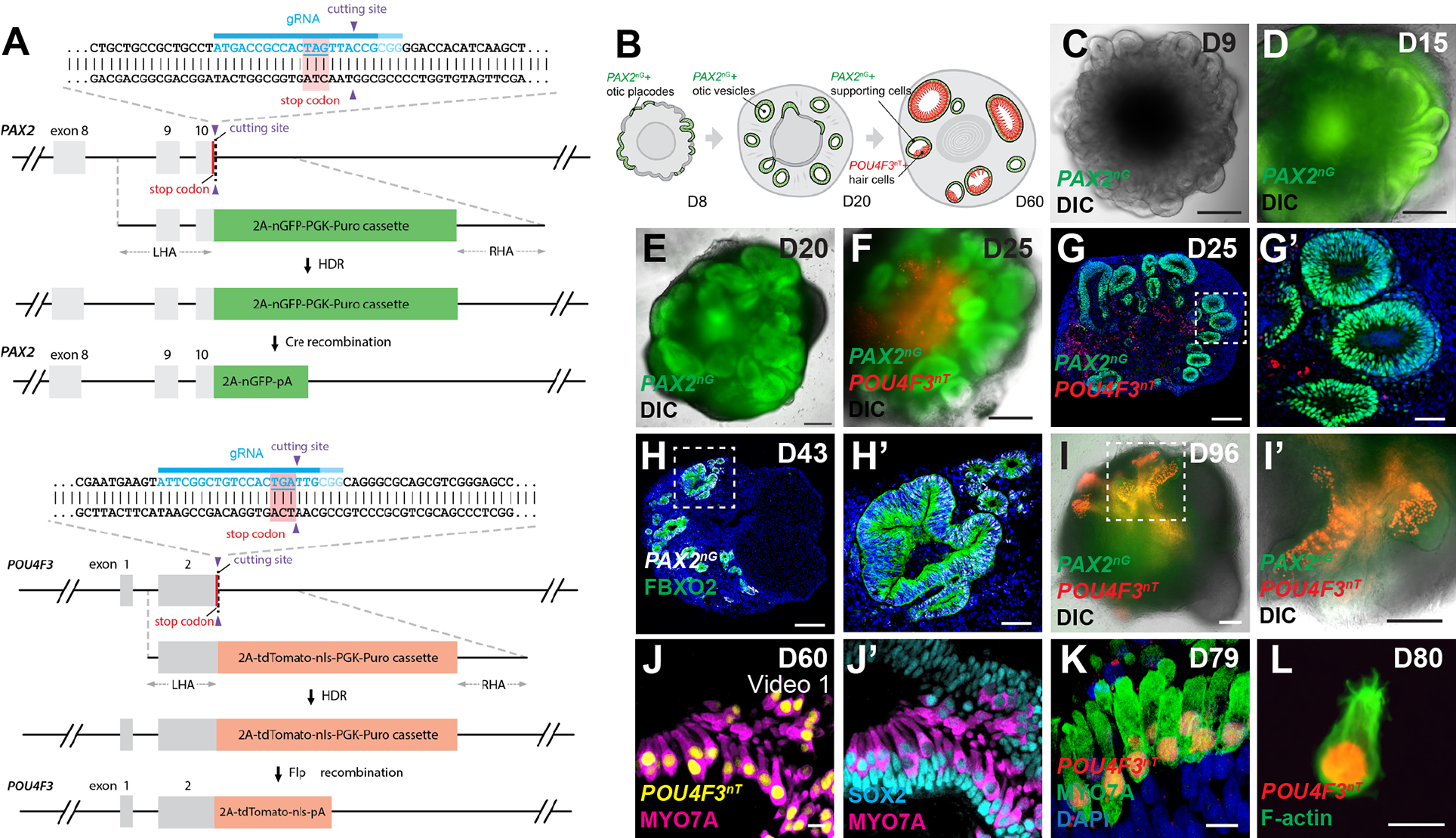
Figure 1. PAX2-2A-nGFP/POU4F3-2A-ntTomato (PAX2nG/POU4F3nT) multiplex reporter hESCs faithfully recapitulate otic progenitor and hair cell differentiations in inner ear organoids.

(A) Schematic illustrations of the PAX2-2A-nGFP and POU4F3-2A-ntdTomato CRISPR design.
(B) Schematic of PAX2nG and POU4F3nT reporter expression during inner ear organoid development.
(C-F) Live images of whole aggregates containing multiple developing inner ear organoids show the spatio-temporal progression of PAX2nG reporter expression and early morphogenesis of PAX2+ epithelium.
(G-H) Representative images of hESC-derived aggregates showing PAX2nG+ epithelium organized into vesicles that co-express the otic-specific marker FBXO2, but devoid of POU4F3nT expression.
(I-I’) Live images of late-stage (D96) aggregates showing intense POU4F3nT+ puncta localized to epithelial vesicles.
(J-K) POU4F3nT+ cells in inner ear organoids also express the hair cell markers MYO7A and SOX2, and are located on the luminal surface of SOX2+ supporting epithelia.
(L) Fixed cell suspension of dissociated POU4F3nT+ cells isolated from D80 inner ear organoids reveals tdTomato+ nuclei and F-actin+ membranes and stereocilia.
Scale bars, 200 μm (C-G, H, I, I’), 50 μm (G’, H’), 10 μm (J-L).
