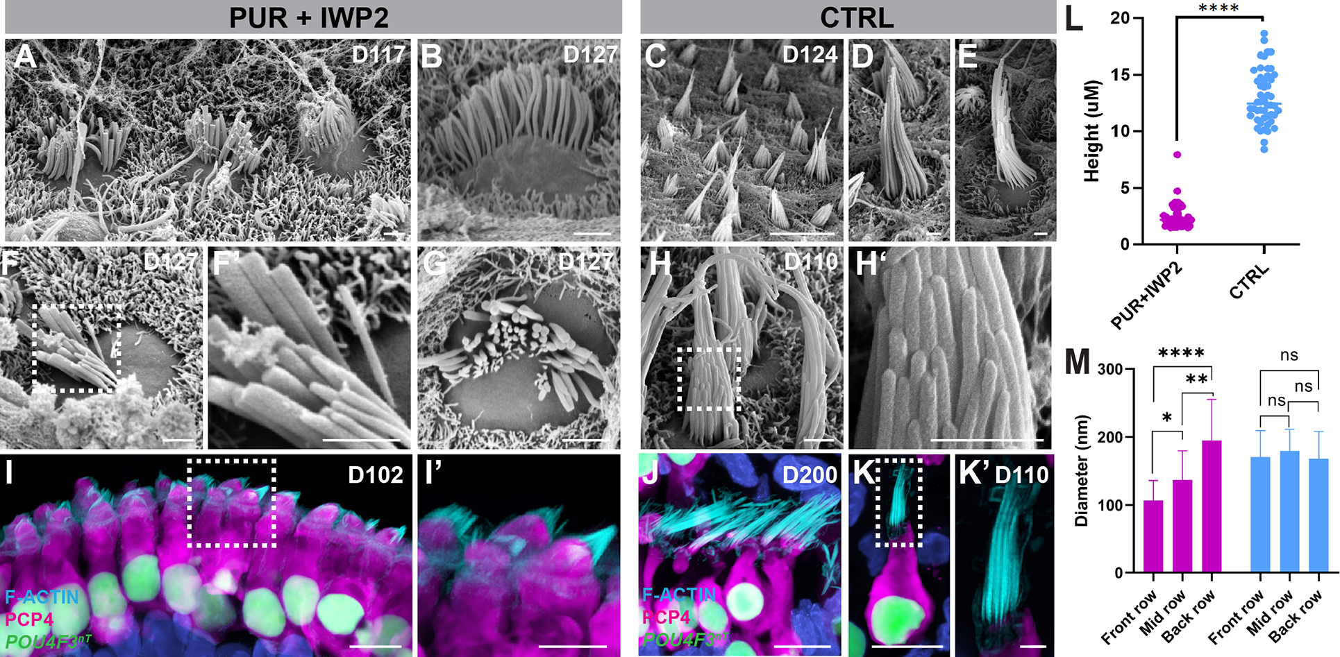
Figure 6. Hair cells derived from ventralized inner ear organoids exhibit structural properties of cochlear hair cells.

(A-H’) Scanning electron micrographs of PUR+IWP2 hair bundles (A-B, F-G) reveal relatively short stereocilia organized into concave rows of increasing height and diameter characteristic of a cochlear hair cell phenotype. In contrast, scanning electron micrographs of CTRL hair bundles (C-E, H-H’) reveal elongated stereocilia organized into convex rows that are of equivalent diameter characteristic of native vestibular hair cells. i-K’, Confocal microscopic images of PUR+IWP2-treated hair cells (I-I’) reveal short F-actin+ hair bundles and rectangular soma with basally-positioned nuclei, whereas those of CTRL hair cells.
(J-K’) reveal elongated F-actin+ hair bundles and an often-bulbous or flask-shaped soma. CTRL hair cells retain vestibular morphology even at D200 (J).
(L-M) Quantitative analysis of the stereocilia height and the diameter of individual stereocilia in PUR+IWP2 vs. CTRL hair cells; n = 50 biological samples from separate experiments: L, Welch’s two-sided t-test; M, One-way ANOVA with Tukey’ ****P<0.00001; values are mean ± SEM.
Scale bars, 10 μm (C, I, J, K), 1 μm (A, B, D-H, K’).
