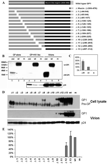
FIG. 1.
Deletion analysis of GP1 in Ebola virus entry. (A) Diagrams of wild-type GP and deletion mutants Δmucin and Δ1 to Δ13. The residues are numbered with the signal peptide (residues 1 to 32) according to GP0 of Ebola virus Zaire. (B) Expression and virion incorporation of Δmucin protein. GP alone, the Δmucin or wt GP gene was used in transfection. GP+HIV Vec, the GP gene and HIV vector were used in transfection. Lysates derived from transfected 293T cells were subjected to SDS-PAGE and Western blotting to detect wt or Δmucin GP1 proteins. Virions, Δmucin or wt GP proteins associated with HIV particles were detected by Western blotting. HIV p24 protein was detected in cell lysates or in the HIV particles. m, mock-transfected cells. (C) The infectivity of Δmucin GP-pseudotyped virus was measured by luciferase assay and is expressed as a percentage of wt infectivity (100%). Error bars indicate standard deviations. (D) Expression and virion incorporation of deletion mutants Δ1 to Δ13. (E) Infectivity of Δ1 to Δ13 GP-pseudotyped virus by luciferase assay.

