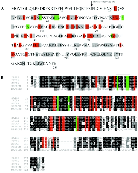
FIG. 7.
Roles of individual residues of GP1 in protein structure and function of filoviruses. (A) Summary of functional analysis of individual residues of Ebola virus GP1. Red, residues involved in protein structure; green, putative receptor-binding residues; grey, no detectable defect in protein structure and function. The putative receptor-binding pocket is highlighted by a line on the top. (B) Sequence alignment of the receptor-binding domains of Ebola virus GP1 and Marburg virus GP1. Residues in red and green are implicated in protein folding and in receptor binding, respectively.

