
Figure 2:
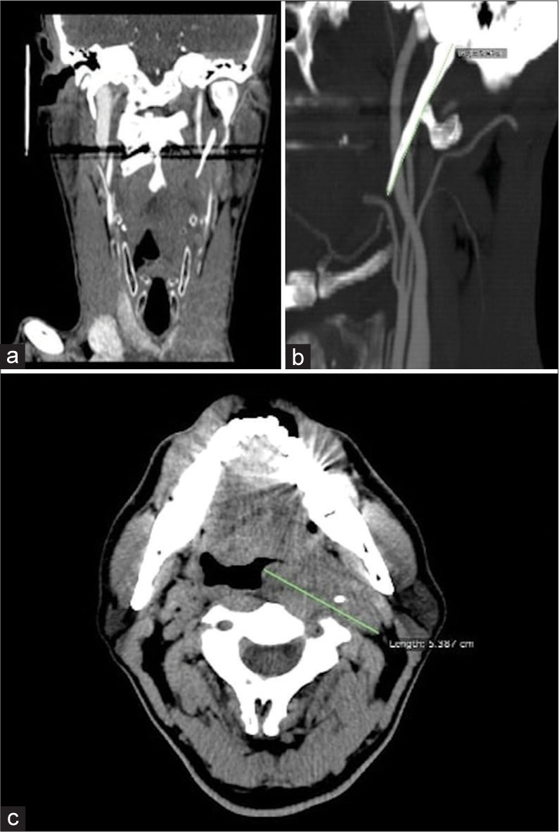
(a and b) Coronal and sagittal computed tomography (CT) scans show the elongated left styloid process causing internal carotid artery compression. (c) The axial CT scan showed hypodense tissue in the left retropharyngeal space with poor enhancement and diameters of about 5 × 3 cm.