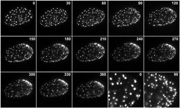
FIG. 3.
Time-lapse live-cell microscopy detecting the redistribution of Sp100 during the initial stages of HSV-1 infection. An uninfected cell expressing EYFP-Sp100 at the edge of a developing HSV-1 dl0C4 plaque was examined by image capture every 15 min. Selected frames from the full sequence are shown, with the times after initiation of the sequence indicated in minutes. The last two images in the third row illustrate expanded views of the right-hand part of the nucleus at the 0- and 90-min time points to demonstrate the increase in the total numbers of ND10-like foci. Selected frames showing the signal from ECFP-ICP4 expressed by the incoming virus are presented in Fig. 4.

