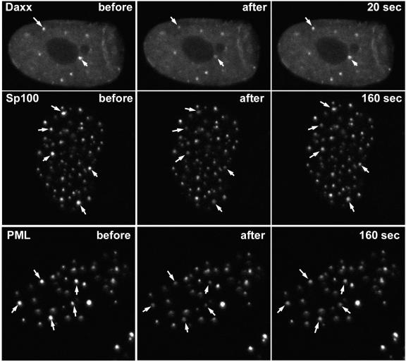
FIG. 6.
Analysis of dynamics of hDaxx, PML, and Sp100 by FRAP. (Top row) HFFF-2 cells were infected with Ac.CMV.EYFP-Daxx, and then the following day, the cells were examined by live-cell confocal microscopy. Two foci of hDaxx (arrows) were bleached by 20 reiterations of the 510-nm laser at full power, and then images were captured at 20-s intervals thereafter. The images before bleaching, immediately after bleaching, and at the first 20-s time point are shown. (Middle row) HFFF-2 cells were infected with Ac.CMV.EYFP-Sp100, and then the following day, the cells were examined by live-cell confocal microscopy. Five foci of Sp100 (arrows) were bleached by 20 reiterations of the 510-nm laser at full power, and then images were captured at 20-s intervals thereafter. The images before bleaching, immediately after bleaching, and at the 160-s time point are shown. (Bottom row) HFFF-2 cells were infected with Ac.CMV.EYFP-PML, and then the following day, the cells were examined by live-cell confocal microscopy. Five foci of PML (arrows) were bleached by 20 reiterations of the 510-nm laser at full power, and then images were captured at 20-s intervals thereafter. The images before bleaching, immediately after bleaching, and at the 160-s time point are shown.

