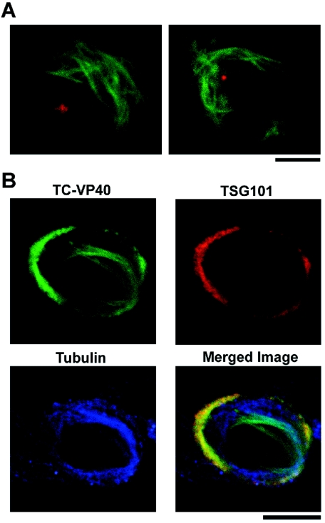
FIG. 2.
VP40 arranged along microtubules does not associate with centrosomes or TSG101. (A) TC-VP401-317 was transfected into 293T cells and stained with FlAsH (green). Cells were subsequently fixed and immunostained with an antibody to γ-tubulin to mark the location of centrosomes. (B) 293T cells expressing full-length TC-VP40 were stained with FlAsH (green) and immunostained with antibodies to TSG101 (red) and α-tubulin (blue). As seen in the merged image, fractions of VP40 colocalized with TSG101 or with microtubules, but little or no colocalization occurred among all three. Bars, 10 μm.

