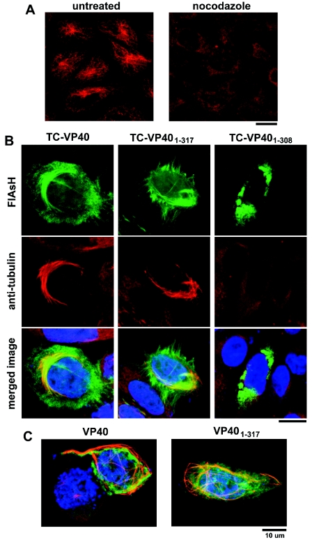
FIG. 5.
VP40 stabilizes microtubules against nocodazole-induced depolymerization. (A) 293T cells immunostained for α-tubulin demonstrate the effect of a 2-h incubation in nocodazole (right) compared to untreated cells (left). (B) Nocodazole (2 μM) was applied to 293T cells transfected with TC-VP401-317 (middle) or TC-VP401-308 (right) for 2 h before fixation. VP40 was visualized with FlAsH (green), and after fixation, the cells were immunolabeled for α-tubulin (red) and stained with Hoechst dye to show cell nuclei. Microtubules are retained in cells expressing full-length VP40 or VP401-317 but not VP401-308. (C) Microtubules can be seen in abundance in cells expressing VP40 or VP401-317 even when cells are treated continuously with nocodazole from within 1 h after transfection. The panel for full-length TC-VP40 also contains a nonexpressing cell for comparison. Bars, 10 μm.

