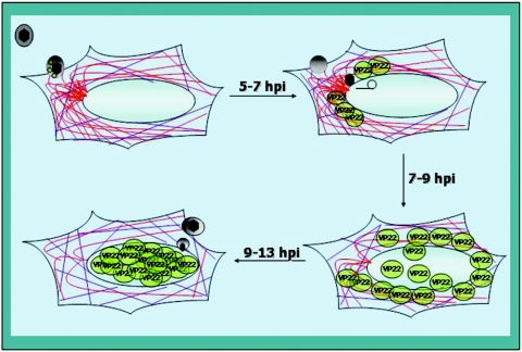
FIG. 9.
Schematic representation of a model for microtubule reorganization and the nuclear localization of VP22 during HSV-1 infection in Vero cells. VP22 (green spheres) and other tegument proteins (gray sphere), including VP13/14, vhs, and VP16, are components of the incoming virion. At times prior to 5 hpi, dense MTOCs and both acetylated (purple) and nonacetylated (red) microtubules are observed in infected cells. The earliest detected VP22 is cytoplasmic and appears to accumulate at perinuclear-juxtanuclear locations, sometimes colocalizing with microtubules (approximately 7 hpi). As VP22 begins to be detected in nuclei, MTOCs decrease (approximately 9 hpi) until they completely disappear (approximately 13 hpi). At 13 hpi, essentially all VP22 observed is localized in nuclei. Throughout this process, acetylated microtubules remain intact.

