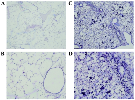
FIG. 4.
Immunohistochemical staining of lung sections at 4 days postinfection. CBA mice were infected intranasally with 4 × 10⁵ PFU of KyA (A and B) or KyAgp2F (C and D). On day 4 postinfection, the mice were sacrificed and the lungs were infused with OCT, removed, and sectioned. The sections were fixed for 20 min in an ice-cold mixture of 75% acetone-25% ethanol, stained with antibody specific for Mac-1, and counterstained with a solution containing 1% methyl green as described in Materials and Methods. Resulting slides were visualized and photographed at 4× (A and C) or 10× (B and D) magnification.

