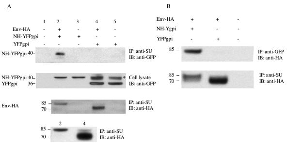
FIG. 4.
MLV NH forms a hetero-oligomer with MLV envelope protein and blocks its proteolytic processing. (A) Coimmunoprecipitation of NH-YFPgpi by anti-SU antibody. HEK293 cells were cotransfected with 1.5 μg each of expression vectors for HA-tagged fusogenic MLV envelope protein (Env-HA) and NH-YFPgpi (or YFPgpi as a control) as indicated above each lane. Twenty-four hours later, the cells were lysed for immunoprecipitation with anti-MLV SU goat serum and blotted with anti-GFP (top panel) or anti-HA (third panel). Samples corresponding to those in lanes 2 and 4 in the top three panels but from an independent transfection are shown in the fourth panel with a longer gel run. Cell lysate before immunoprecipitation was blotted with anti-GFP antibody as a control (second panel). Two species of YFPgpi were detected with the anti-GFP antibody, one of which migrated anomalously slowly and is marked with * in the second panel. (B) Coimmunoprecipitation of envelope protein by anti-GFP antibody. The same procedures as described for panel A were carried out except that anti-GFP antibody was used for immunoprecipitation instead of anti-SU goat serum. Molecular mass markers (in kilodaltons) are shown at the left.

