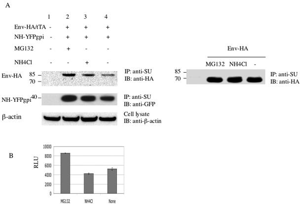
FIG. 6.
MLV NH slightly increases the level of degradation of MLV envelope protein, mainly through the proteasome pathway. (A) Two micrograms each of expression vectors for Env-HA and tTA were cotransfected into HEK293 cells with (left) or without (right) NH-YFPgpi (2 μg). Twenty-four hours later, these cells were treated with the proteasome inhibitor MG132 (20 μM), treated with the lysosomal degradation inhibitor NH4Cl (20 mM), or not treated. After 10 h, cells were lysed, and the lysate was immunoprecipitated with anti-SU goat serum and blotted with anti-HA (top left and right) or anti-GFP (middle left) antibody. An aliquot of cell lysate that was not immunoprecipitated was blotted with anti-β-actin antibody (bottom left) as a control. Molecular mass markers (in kilodaltons) are shown at the left. IP, immunoprecipitation; IB, immunoblotting. (B) Transfected HEK293 cells corresponding to lanes 2 to 4 in panel A were cocultivated with U2OSLucCATGFP cells and assayed for luciferase activity. The x-axis label indicates inhibitor treatment. Error bars indicate the standard deviations of results with triplicate samples in a representative experiment of at least three independent transfections. RLU, relative light units.

