
Figure 1.
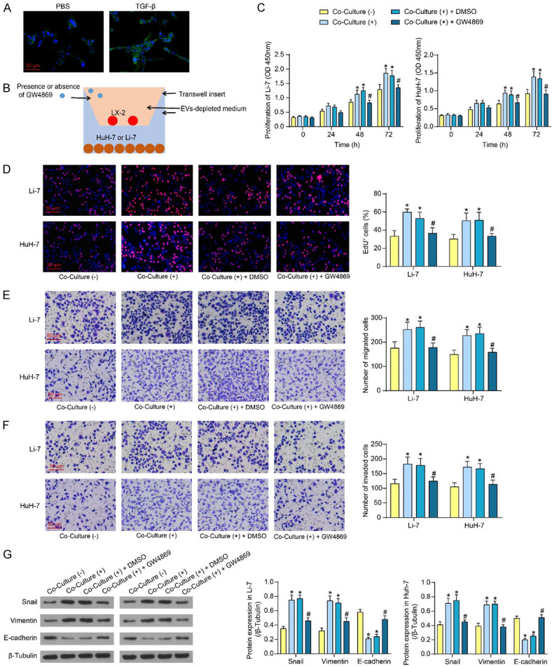
Blocking the secretion of EVs by LX-2 inhibits its effects on HCC cell biological behavior. A. Expression of activation marker protein α-SMA after TGF-β (2 ng/mL) treatment of LX-2 cells for 36 h detected by immunofluorescence. B. Schematic diagram of co-culture of activated HSC with HCC cells. C. The proliferative activity of HCC cells after co-culture with LX-2 in the presence or absence of GW4869 was measured using CCK8. D. EdU+ cells in HCC cells after co-culture with LX-2 in the presence or absence of GW4869 were measured using EdU staining. E. The migratory activity of HCC cells after co-culture with LX-2 in the presence or absence of GW4869 was measured using Transwell assay. F. The invasive activity of HCC cells after co-culture with LX-2 in the presence or absence of GW4869 was measured using Transwell assay. G. EMT-related protein expression in HCC cells after co-culture with LX-2 in the presence or absence of GW4869 was measured using western blot assays. The results represent means ± SD, *P < 0.05 vs Co-Culture (-); #P < 0.05 vs Co-Culture (+) + DMSO. All experiments were repeated at least three times, two-way ANOVA.