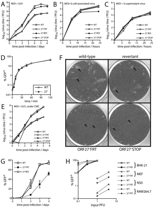
FIG. 5.
ORF27 mutant viruses showed impaired low-multiplicity growth in BHK-21 cells. (A) BHK-21 cells were infected at a low multiplicity (0.01 PFU/cell) with ORF27+ (WT and REV) and ORF27− (FRT and STOP) viruses as indicated. The amounts of infectious virus in cultures were then determined by a plaque assay at the indicated times. Data for one of three equivalent experiments are shown. (B) BHK-21 cells were infected at a high multiplicity (5 PFU/cell) for 2 h at 37°C. The input virus was removed by acid washing, and the infectious virus associated with the cells was measured by a plaque assay as described for panel A. (C) After high-multiplicity infection and acid washing, the amount of infectious virus released into cell-free supernatants over time was measured by a plaque assay. (D) BHK-21 cells were exposed to the GFP+ wild-type (WT) or ORF27− FRT (27−) virus (1 PFU/cell) at 37°C for the indicated times. The cells were then acid-washed and fed with new medium. Sixteen hours later, the proportion of GFP+ cells was determined by flow cytometry. GFP expression occurred by virtue of retaining the loxP-flanked BAC cassette at the left end of the viral genome. (E) BHK-21 cells were infected at a low multiplicity as described for panel A but were then cultured in medium with 0.6% carboxymethylcellulose (CMC) to prevent the spread of virions throughout the culture medium. (F) BHK-21 monolayers were infected with the indicated viruses and cultured under 0.6% carboxymethylcellulose. The arrows show example plaques. At the dilutions shown, there were larger plaque numbers on the monolayers infected with the ORF27 mutant viruses, but the plaques were smaller. (G) BHK-21 cells were infected with the GFP+ wild-type (WT), ORF27-deficient (27− FRT), or revertant (27 REV) virus as indicated, at a multiplicity of 0.01 PFU/cell. Replicate cultures were then maintained in medium supplemented with 2% serum pooled from MHV-68-immune mice. At the time points indicated, GFP expression in sample cultures was determined by flow cytometry of trypsinized cells. Each point shows the mean ± standard deviation of triplicate cultures. (H) BHK-21 cells, MEFs, NS0 myeloma cells, and RAW264.7 macrophages were infected with the GFP+ wild-type (WT) or ORF27-deficient (27− FRT) virus, as indicated. Eighteen hours later, the cells were analyzed for GFP expression by flow cytometry. PFU titers of virus stocks were determined on BHK-21 cells. The data shown are representative of three independent experiments.

