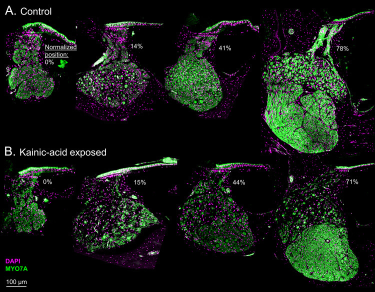
Fig. 2.
Comparison between representative control (A) and kainic acid (KA)-exposed (B) frozen cross-sections at different cochlear locations (marked below the HC epithelium in each panel). Normalized position is the percent distance from the apical to the basal end of the cochlear ganglion. Note that there are no ganglion cells in the left column because the ganglion does not extend to the extreme apical end of the HC epithelium. KA visibly reduces density of ganglion cells and distal AN fibers without impacting the HC epithelium

