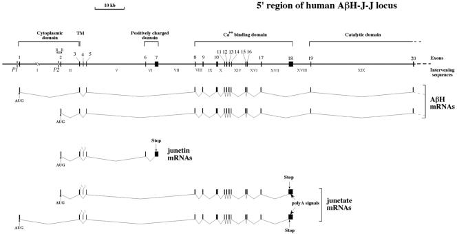
FIG. 1.
Structure of the 5′ end of the human locus for aspartyl-β-hydroxylase, junctin, and junctate. Arabic numbers over black boxes indicate exons. Intervening sequences are indicated in roman numerals. The two putative promoters P1 and P2 are indicated. A schematic representation of aspartyl-β-hydroxylase, junctin, and junctate exon splicing (11, 60) is shown at the bottom of the panel. The cytoplasmic, transmembrane (TM), positively charged, calcium binding, and catalytic domains are indicated. The locations of AUG, stop codons, and poly(A) signals are shown. The BglII (B) plasmid subclone of BAC 1 (60) covering the second exon of the locus is also shown.

