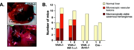
FIG. 2.
Inhibition of vascular-tumor development in Vhlh/Arnt mutant mice. (A) Photographs of gross cavernous liver hemangiomas observed in PEPCK-Vhlh and PEPCK-Vhlh/Hif-1α mutant mice. Hemangiomas are indicated by arrows. (B) Incidence of macroscopic liver hemangiomas and microscopic vascular lesions observed in PEPCK-Cre mutant mice. Mice from the indicated genotypes were divided into two age groups for analysis, mice 2 to 6 months of age (group 1) and mice >6 months of age (group 2). Each bar represents the number of mice from the indicated genotype with macroscopic hemangiomas, microscopic vascular lesions in the liver, and normal liver. A chi-square test revealed that there were no significant differences between the distributions of macroscopic and microscopic liver hemangiomas in the Vhlh- and Vhlh/Hif-1α-deficient mice.

