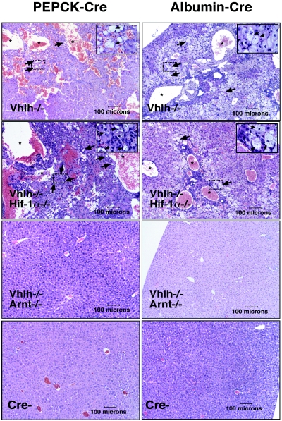
FIG. 3.
Inactivation of Arnt suppresses Vhlh-associated vascular lesions and steatosis. Hematoxylin and eosin staining of PEPCK-Cre (left; >6 months of age) and Albumin-Cre (right; 4 to 6 weeks of age) mutant liver sections (magnification, ×100). Note that large vascular spaces (stars) and steatosis (arrows) were observed in both Vhlh and Vhlh/Hif-1α mutant mice. The black boxes outline areas that are shown at higher magnification (×1,000) in the upper right corners.

