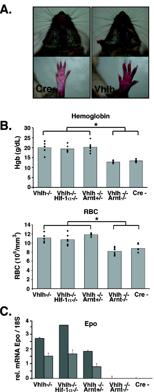
FIG. 4.
Inactivation of Vhlh and Vhlh/Hif-1α in hepatocytes induces erythrocytosis. (A) Control (Cre−) and PEPCK-Vhlh muzzles and paws. Note the increased red coloration of the skin in the PEPCK-Vhlh mutant mouse. (B) Elevated hemoglobin and red blood cell numbers in PEPCK-Vhlh- and PEPCK-Vhlh/Hif-1α-deficient mutant mice. The results shown are average hemoglobin concentrations and RBC numbers in blood collected from PEPCK-Cre mutant mice determined by a CBC analyzer. Note that Vhlh/Arnt mutant mice and control mice have similar hemoglobin concentrations and red blood cell numbers (no statistical differences as determined by t test). Vhlh-, Vhlh−/−/Hif-1α−/−, and Vhlh−/−/Arnt+/− mice exhibited increased levels of hemoglobin and red blood cells compared to control and Vhlh−/−/Arnt−/− mice (*, P < 0.05). Six mice from each genotype were analyzed and are represented by individual dots. (C) Liver erythropoietin expression correlates with erythrocytosis. Shown are Epo mRNA expression levels from PEPCK-Cre mutant livers determined by real-timePCR. Each bar represents the average of three values obtained for an individual mouse. The error bars represent standard deviations. 18S was used to normalize mRNA. Two representative mice from an n of 3 are shown for each genotype.

