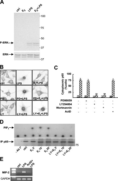
FIG. 6.
A nongenomic, PI3K-dependent pathway is necessary for the action of E2. (A) Western blot analysis of phosphorylated ERK (P-ERK) and ERK proteins in total extracts from RAW 264.7 cells treated with 1 nM E2 for 10 min before treatment with LPS at 50 μg/ml for 30 min. ctrl, control. (B) Subcellular localization of p65. Immunocytochemical analysis with anti-p65 antibody was performed on RAW 264.7 cells treated with PD98059 (50 μM) (PD) or LY294002 (50 μM) (LY) for 1 h, with 1 nM E2 for 10 min, and with LPS at 50 μg/ml for 30 min as indicated. (C) Quantification of E2 activity shown in panel B. The percentage of cells with cytoplasmic p65 is plotted relative to the total cell number. Black bars, LPS; hatched bars, E2 plus LPS. Actinomycin D (5 μg/ml) (ActD) was added 1 h before E2 and LPS. Values are the means ± SDs of a single experiment performed in triplicate. Single and double asterisks indicate P values of <0.05 in a comparison with LPS and in a comparison with E2 plus LPS, respectively. (D) PI3K activity. RAW 264.7 cells were treated in the presence or absence of 50 μM LY294002 for 1 h and then with 1 nM E2 for 5, 10, or 20 min. Cell lysates were immunoprecipitated with anti-p85 antibody (lower panel), and PI3K activity was assayed. The upper panel shows a representative autoradiogram of a thin-layer chromatography analysis of the migration of labeled PIP3. (E) Gene transcription inhibition by actinomycin D. MIP-2 mRNA levels in RAW 264.7 cells treated with actinomycin D (5 μg/ml) for 1 h and then with LPS for 1 h were assessed by RT-PCR.

