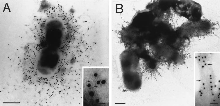
FIG. 5.
Analysis of SEF14 and SEF21 fimbria assembly in S. enteritidis 3b-122 pLU/TA 4-1 by immunogold electron microscopy. S. enteritidis 3b-122 pLU/TA 4-1 was labeled with protein A-gold and negatively stained following incubation with immune serum generated to SEF14 (A) or SEF21 (B). Arrows indicate individual immunogold-labeled SEF14 and SEF21 fimbriae in panel A and B insets, respectively. The average diameter of the gold particles was 15 nm. Bar, 0.5 μm (electron micrograph) or 50 nm (inset).

