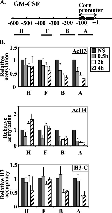
FIG. 5.
Histone proteins are lost across the GM-CSF promoter after EL-4 T-cell activation. (A) Diagram of the GM-CSF gene promoter showing the transcription start site (+1) and the defined core promoter region. The regions amplified by GM-CSF PCR primer sets are shown by the lines labeled with capital letters under the map. The sequences of the GM-CSF PCR primer sets are shown in Table 1. (B) EL-4 cells were left nonstimulated (NS) or stimulated with P/I for 30 min, 2 h, or 4 h, and ChIP assays were performed with antibodies directed against acetylated H3 (AcH3), acetylated-H4 (AcH4), and the C terminus of H3 (H3-C). The immunoprecipitated DNA was amplified with the primer sets shown. The data are shown relative to total input DNA and normalized to the value for nonstimulated cells for each GM-CSF primer set. The values represented the combined means ± standard errors (error bars) of three independent experiments.

