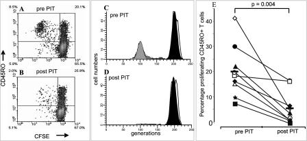
Figure 1. PIT Reduces Antigen-Specific Proliferation of Memory T Cells.
(A–D) PBMCs taken before and after PIT were labelled with CFSE to track cell division after antigen stimulaton. Proliferation of cat-allergen-specific CD45RO+ lymphocytes was reduced following PIT (A) and (B). (C) and (D) represent CD45RO+ T cells as shown in panels (A) and (B), respectively, analysed with ModFit software. The right-hand peaks represent the parental population, and generations of dividing cells are depicted leftwards along the x-axis.
(E) Summary of the percentage of proliferating CD45RO+ T cells pre- and post-PIT (percent proliferating cells is defined as the fraction of the starting population that has proliferated during the course of the experiment, determined with Modfit) for all nine patients tested. Open symbols represent patients enrolled in treatment Group 1, while solid symbols depict patients from treatment Group 2. Horizontal solid bars show mean levels of proliferation. Background proliferation (in the absence of a stimulus) was less than 2% and was subtracted. The Wilcoxon signed rank test was used for statistical analysis.

