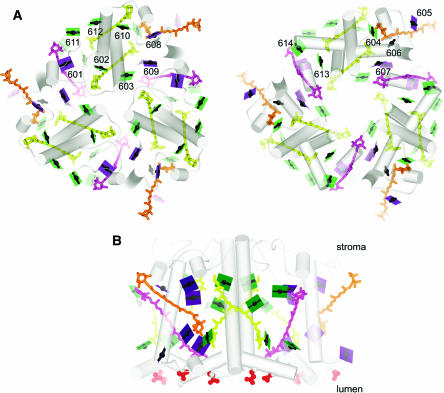
Figure 3.
Organization of Pigments in LHCII.
(A) The trimer is viewed along the membrane normal from the stromal side (left). The lumenal side (right) is viewed from the stromal side as if reflected in a mirror to facilitate visualization of chlorophyll proximities in the two layers. The color schemes of green for chlorophyll a, purple for chlorophyll b, the arbitrary numbering system for the pigments from 601 to 614, and representation of the chlorophylls as a line connecting two nitrogens of rings A and C with a central magnesium (see Figure 4), which emphasizes the direction of the Qy axis, is retained from Liu et al. (2004). The carotenoids are indicated in yellow for luteins, orange for neoxanthin, and magenta for the xanthophyll cycle pigment. As in Figure 2, the pigments closer to the viewer appear darker than those further away. Hence, 24 chlorophylls are shown as dark planes in the stromal view. The lighter ones in that image are the ones in the lumen-side layer.
(B) The trimer is viewed in the plane of the membrane, perpendicular to the membrane normal as in Figure 2. The red spheres show the negatively charged residues on the lumen side.

