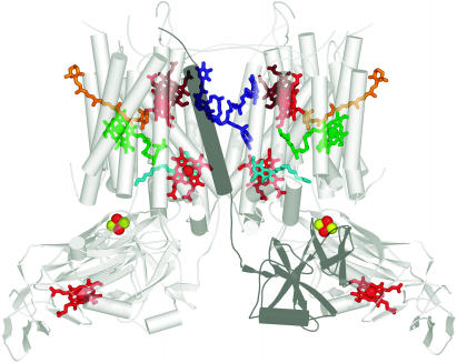
Figure 5.
Cofactors in the b6f Dimer.
The coordinates from the cyanobacterial structure were used to generate the figure (Kurisu et al., 2003). The complex is viewed in dimeric form in the plane of the membrane with the lumen side on the bottom as in Figure 2. Chlorophyll is shown in green, b hemes of cytochrome b6 and the c-heme of cytochrome f in red, the novel heme in brown, and Fe and S as red and yellow balls, respectively. The Qo site inhibitor, tridecyl stigmatellin, is shown in turquoise and a plastoquinone at the Qi site in blue. One of the Rieske subunits is shaded as a dark gray to show the association of its cofactor-containing domain with the other monomer. The stromal side of the membrane is called the n-side in one work or the inside (i) in another, and the lumen side is called p-side or outside (o). Allen (2004) has discussed the variety in nomenclature of cofactors and presentation of the complex.

