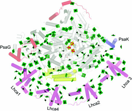
Figure 6.
Association of LHCI with PSI.
The complex is viewed along the membrane normal from the stromal side. The chlorophylls are shown as green planes with the central magnesium as a green sphere. Fe is shown as a red sphere and sulfur as a yellow sphere. The helices are shown as cylinders. The subunits of PSI are colored gray, with the exception that polypeptides novel to the plant PSI are colored pale red (top left of image) and PsaK is shown in blue and PsaF in yellow (see Scheller et al., 2001 for a review of PSI polypeptides). The Lhca polypeptides are colored pale magenta (bottom of image). PyMOL was used to generate the image from coordinates provided by Adam Ben-Shem and Nathan Nelson (Ben-Shem et al., 2003).

