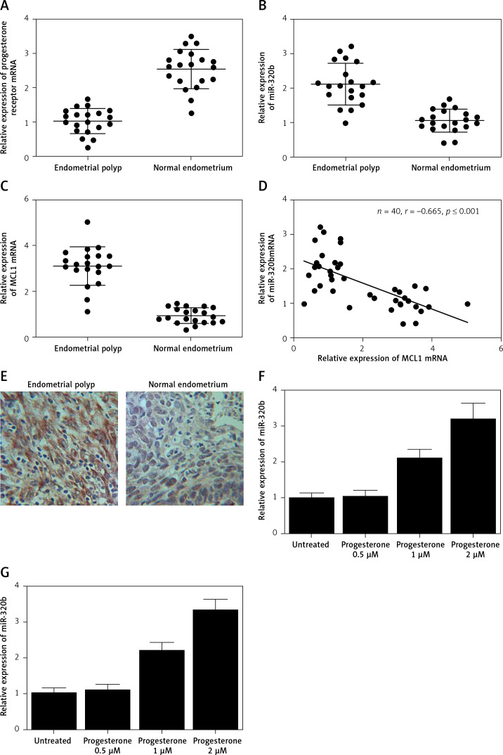
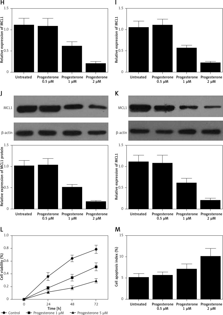
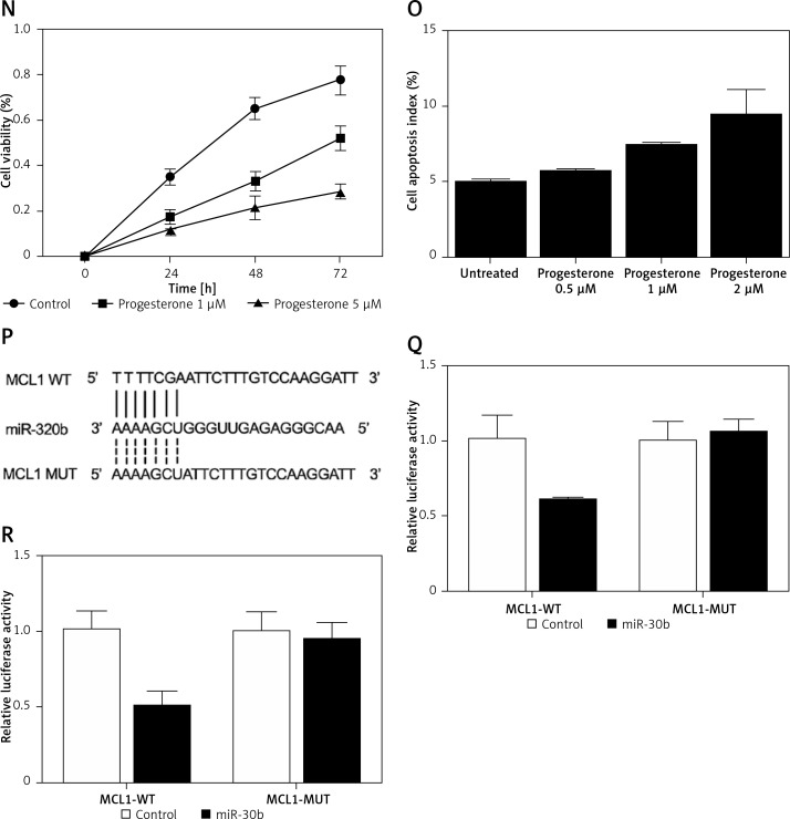
Figure 1.
A – mRNA level of progesterone receptor in the EP group was much lower than that in the control group. B – MiR-320b expression in the EP group was much lower than that in the control group. C – mRNA level of MCL1 in the EP group was much lower than that in the control group. D – A negative regulatory relationship with a correlation coefficient of –0.5000 was confirmed between miR-320b and MCL1 expression. E – The protein level of MCL1 in the EP group was much higher than that in the control group. F – 0.5 μM of progesterone exerted no effect on the expression of miR-320b, while 1 and 2 μM of progesterone increased miR-320b expression in RL95-2 cells in a dose-dependent manner. G – 0.5 μM of progesterone exerted no effect on the expression of miR-320b, while 1 and 2 μM of progesterone increased miR-320b expression in HEC-1-A cells in a dose-dependent manner. H – 0.5 μM of progesterone exerted no effect on the expression of MCL1 mRNA, while 1 and 2 μM of progesterone decreased MCL1 mRNA expression in RL95-2 cells in a dose-dependent manner. I – 0.5 μM of progesterone exerted no effect on the expression of MCL1 mRNA, while 1 and 2 μM of progesterone decreased MCL1 mRNA expression in HEC-1-A cells in a dose-dependent manner. J – 0.5 μM of progesterone exerted no effect on the expression of MCL1 protein, while 1 and 2 μM of progesterone decreased MCL1 protein expression in RL95-2 cells in a dose-dependent manner. K – 0.5 μM of progesterone exerted no effect on the expression of MCL1 protein, while 1 and 2 μM of progesterone decreased MCL1 protein expression in HEC-1-A cells in a dose-dependent manner. L – Progesterone (1, 2 μM) dose-dependently inhibited the proliferation of RL95-2 cells. M – Progesterone (1, 2 μM) dose-dependently inhibited the proliferation of HEC-1-A cells. N – Progesterone (1, 2 μM) dose-dependently promoted the apoptosis of RL95-2 cells. O – Progesterone (1, 2 μM) dose-dependently promoted the apoptosis of HEC-1-A cells. P – MCL1 was identified as a target gene of miR-320b with a ‘seed sequence’ located in the 3’UTR of MCL1. Q – Luciferase activity of RL95-2 cells co-transfected with miR-320b mimics and wild-type MCL1 3’UTR was much lower than that of the cells co-transfected with miR-320b mimics and a scramble control. R – Luciferase activity of HEC-1-A cells co-transfected with miR-320b mimics and wild-type MCL1 3’UTR was much lower than that of the cells co-transfected with miR-320b mimics and a scramble control


