Figure 6.
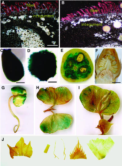
Histochemical Localization of GUS Activity in Transgenic Cotton Plants Containing the GhACT1∷GUS Fusion Genes.
(A) and (B) Dark-field micrographs of 5-μm-thick cross sections of 1- to 2-DPA ovules. A high level of GUS activity (represented by pink dots) was only found in the fiber, and very weak GUS staining was seen in the inner cell layers. No GUS staining was detected in the epidermal atrichoblast and integument.
(C) to (J) Bright field of micrographs or photographs of ovules and other tissues/organs.
(C) and (D) GUS staining in ovules at 1 (C) and 2 (D) DPA. Strong GUS activity was observed in the fibers.
(E) A cross section of a transgenic cotton boll at 14 DPA. Strong GUS activity was detected in the developing fibers, and very weak GUS staining was seen in embryos.
(F) A longitudinal section of a transgenic flower bud before anthesis. Weak GUS staining was found in some pollen grains.
(G) to (I) GUS staining in transgenic seedlings.
(G) Three-day-old seedling. GUS gene was expressed moderately in the cotyledons.
(H) Parts of a 7-d-old seedling. Weak GUS expression was found only in cotyledons.
(I) Parts of a 10-d-old seedling. GUS activity was very low in cotyledons, and no GUS expression was detected in other tissues, such as leaf and shoot apex.
(J) No GUS activity was detected in leaf, stem, root, sepal, and petal (from left to right) of transgenic cotton.
Bars = 80 μm in (A) and (B), 1 mm in (C) and (D), and 2 mm in (F).

