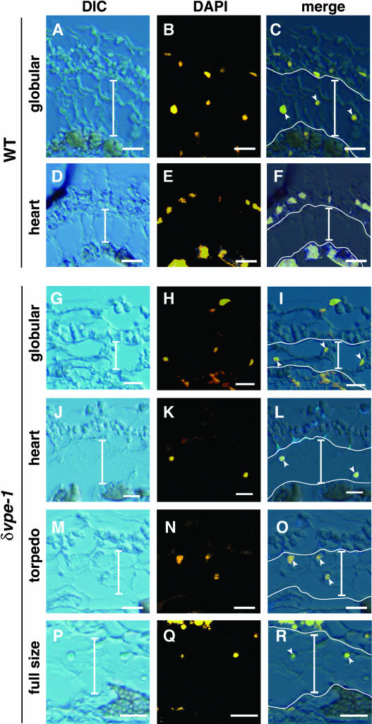
Figure 5.
Degradation of Nuclei in ii2 and ii3 Cell Layers of the Inner Integuments Occurs in the Wild-Type Seeds at the Heart-Shaped-Embryo Stage, whereas It Does Not Occur in the δvpe-1 Seeds at the Early Stages.
Differential interference contrast (DIC) images, DAPI images, and merged images (merge) of developing seeds from the wild type ([A] to [F]) and δvpe-1 ([G] to [R]) are shown. DAPI images were visualized using 365/12-nm-wavelength excitation and a long-pass 397-nm-wavelength emission filter and then were converted in yellow to highlight the nuclei. Developmental stages are indicated at the left. Vertical bars indicate both ii2 and ii3 cell layers. Arrowheads indicate nuclei stainable with DAPI in the ii2 and ii3 cell layers. The cells of ii2 and ii3 layers, which are highly vacuolated, are larger than the cells of the other layers. The cells of outer integument layers contain a lot of starch granules, and the cells of ii1 layer accumulate pigments, which generated autofluorescence. Horizontal bars = 25 μm.

