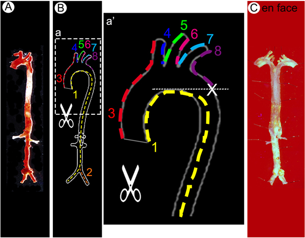
Figure 4.
Step-by-step protocol for en face aorta preparation. (A-B) The arterial tree stained with Oil Red O was opened longitudinally in order to flatten the aorta for imaging. Dotted lines along the vessel wall and numbers indicate sequential cuts to be made to open up the vessels. (C) Longitudinally split and pinned whole aorta on a wax petri dish in Y shape.

